ANATOMY OF THE URETHRA
The helpful key to understanding the medical terminology related to a urethral stricture is to have a basic understanding of both the function and anatomy of the male urethra. The primary function of the urethra is to transport urine from the bladder to the tip of the penis, allowing the bladder to empty when urinating. Normally, the urethra has no restrictions throughout the entire tube, allowing the bladder to empty with an uninterrupted flow. Urethral stricture disease is when the urethra has some kind of blockage or narrowing that restricts the flow of urine.
Although this is often referred to as a “disease”, it is in no way contagious and is rarely associated with any other medical problems. The primary symptom of the disease is obstruction during urination, meaning that as the bladder contracts to empty, the flow through the urethra is impaired. Think of it as something similar to the way a pinch in a garden hose leads to a major reduction in water flow.
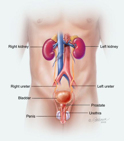
Urine is made in the kidneys, then travels through tubes called ureters to the bladder as shown in the illustration below.
The bladder then fills with urine. Once the bladder is full, the distension of the bladder is sensed as an urge to urinate. During urination, the bladder contracts (squeezes) This contraction continues until the bladder is completely empty. Immediately after urination, there should be no urine or very little residual urine remaining in the bladder.
As the bladder contracts or squeezes, urine first passes through an opening called the bladder neck, a connection between the bladder and the prostate as shown in the illustration below.
The bladder neck is a main area responsible for continence (not leaking urine). The bladder neck is closed most of the time and this keeps the urine in the bladder. However, when the bladder is emptying as a man is urinating, the bladder neck reflexively opens, allowing the urine to pass through.
After the urine gets back the bladder neck, it then travels through the prostate (a gland that surrounds the urethra). This portion of the urethra, closest to the bladder, is called the prostatic urethra.
As a side note, during ejaculation but not urination, secretions from the prostate and other glands called seminal vesicles along the sperm made in the testicles all enter the prostatic urethra which then is propelled down and out the tip of the penis.
As the urine continues to travel on it’s journey towards the penis, it enters a short segment called the membranous urethra, which is surrounded by a muscle called the external urinary sphincter. This is also a source of continence. This external sphincter muscle is generally relaxed only during urination. If I man wants to try to stop during urination mid-stream, what makes that possible is the voluntary contraction of the external sphincter. However, it is generally not a good idea to try to stop urinating in the middle of urination. The membranous urethra is the portion of the urethra often damaged when the pelvic is fractured during a motor vehicle accident or pelvic crush injury. This urethral trauma is called a pelvic fracture urethral injury.
Just a couple of urethra anatomy details – The bladder neck and the external sphincter are two distinct continence mechanisms that prevent involuntary urine leakage (incontinence). As long as one is functional, a man will not be incontinent (leak urine). In a way, it is similar to the main water line and a faucet connected to a garden hose in the home. If the water is turned off at the main water line, then water will not come out of the garden hose, regardless of the setting at the faucet. Moreover, if a working faucet is in the “off” position, water will not exit the hose when the main water line is not turned off. One other detail is that the prostatic urethra and the membranous urethra are together called the posterior urethra.
As the urine travels further on it’s journey towards the penis, it enters the bulbar urethra, the portion of the urethra just below the membranous urethra that is surrounded by the external sphincter area. The bulbar urethra continues until the base of the penis is reached. This portion of the urethra travels under the skin in the area between the scrotum and anus (called the perineum) and within the scrotum between and deep to the testicles. When a man forcefully straddles a fence or a bicycle bar, the bulbar urethra is at risk for injury. Straddle injury is a very common cause of urethral stricture. This is because the urethra in this area is not well protected and is close to the skin. Impact injuries from straddle trauma or a man being kicked crush the urethra and surrounding tissue against the bone, leading to subsequent scarring and an associated narrowing of the urethra.
As the urine continues on towards the tip of the penis, the next area it encounters is the penile urethra. This portion of the urethra courses along the undersurface of the penis. Most men think the major part of the urethra is within the penis, but the bulbar urethra is just as long and more oven involved than the penile urethra. The bulbar and penile urethra are just tubes or hoses. They have nothing to do with continence just like a garden hose is just a way of transporting water from one point to another and it is the faucet that controls the flow.
The head of the penis is called the glans penis. The opening of the urethra at the tip of the glans is called the urethral meatus. The last portion of the urethra after the penile urethra through which the urine travels before reaching the tip (urethral meatus) is the part within the head of the penis. The urethra anatomy term someone gave to this part of the urethra is “fossa navicularis”. Sometimes, male urethra anatomy terms are easy like “penile urethra” but occasionally, there are more complicated words like “fossa navicularis”.


