The first step in diagnosing a urethral stricture or formulating an effective urethral stricture treatment plan is what is getting the patient’s full medical history. The “history” is the patient’s report of his urinary symptoms, a description of any prior treatment, and general information such as medical illnesses, any prior surgeries and urethral dilation or urethrotomy, or urethroplasty surgeries in particular, allergies etc. It is not unusual for our patients to have had failed prior urethral stricture treatment with dilations, incisions, and/or open surgery.
We encourage patients to hand carry their medical records to their initial appointment. This guarantees that the records are available for review at the time of the visit. It is our experience when patients rely on records being faxed from other offices that the records are not received prior the scheduled appointment. When a surgery is performed, it is required that the surgeon dictate a detailed report called the “operative dictation”. This is then transcribed, and it then becomes a part of the hospital chart. We find the operative dictations of prior urethral surgeries most useful.
During the complete physical exam, particular attention should be devoted to assessment of the appearance of the penile skin and the urethral opening. In some cases, an option for urethral reconstruction can be a repair using the penile skin as a “patch”, and when indicated, is performed when a man is uncircumcised with normal skin or is circumcised with some redundancy of the skin (extra skin). The appearance of the urethral opening (called the urethral meatus) is also important. If there is narrowing of the urethral at the tip of the penis, this is called “meatal stenosis”. If there is associated whitish color change of the urethral tip opening, especially when there are also abnormalities of the skin of the penis, this can be diagnostic of Lichen Sclerosis, also called Balanitis Xerotica Obliterans (see section on Lichen Sclerosis).
After the history and physical, then next step is to evaluate the urethra. That is the only way to know what is going on inside the urethra and what is the exact length and location and severity of the urethral stricture.
Bougie Calibration
When the opening of the urethra at the tip of the penis (urethral meatus) is narrow or if there is narrowing just deep to the urethral opening (an area called the “fossa navicularis”), the best way to determine the actual caliber (circumference = size) of the urethra of this portion of the urethra is with instruments called Bougie-a-boules.
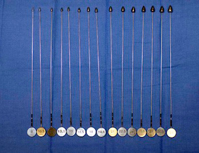
Set of Bougie-a-Boules
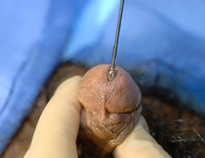
Bougie being used
These cone shaped instruments painlessly measure the circumference of the opening of the urethra the way feeler gauges precisely measure the gap on a spark plug. The measurement of the caliber of the urethra is in French size. French is a measurement of millimeters circumference, and the French size is generally 3 times the diameter. Simply put, the goal is to evaluate the urethra at the tip of the penis to see if there is urethral stenosis there, or if the stricture starts further inside the urethra. A normal adult urethra is greater than 30mm circumference (30 French, approximately 10mm or 1cm diameter) except in the area near the tip of the penis, where the opening is normally approximately 24 French.
When we calibrate the distal (the part closest to the tip) urethra is calibrated, numbing lubricant is placed at the opening of the urethra, and a very small bougie, generally 8 French, is advanced a short distance and then removed. Unless the distal urethra is less than 8 French, the instrument will pass without any resistance. A 9.5 French Bougie is then advanced. We continue the procedure with larger instruments until there is a slight resistance, generally felt as the instrument is removed given that bougies are cone shaped. Once there is very slight resistance, this is the caliber of the urethra. No further instruments are placed as to continue with larger instruments would only dilate/tear the urethra causing pain, and our objective is to painlessly measure the size of the opening. In looking at the pictures of these long metal instruments, one may think the procedure involves jamming these through the penis. However, that is not how they are used (at least at our Center). The instrument is generally advanced only 1 inch after numbing jelly lubricant is placed.
Male Cystoscopy – Urethroscopy
Another part of the diagnostic evaluation for a urethral stricture is a urethroscopy. A urethroscopy is a procedure performed in the office setting. After gently instilling lubricant into the tip of the penis that contains lidocaine, a small (16 French telescope is gently placed into the urethra with advancement to the stricture. This instrument is called a cystoscope or a “penis scope”. It is a flexible tube that has an eye piece at one end and an opening at the opposite tip that allows light to shine through. The procedure is often called cystoscopy, but technically, if the penis scope is just advanced into the urethra and not all the way to the bladder, the procedure is called urethroscopy.
Flexible Scope
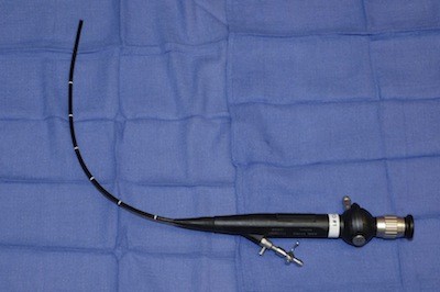
When male cystoscopy is performed, the gentle advancement of this instrument through the tip of the penis allows the Urologist to look inside of the urethra. It is our practice to use a digital cystoscope connected to a high definition monitor so our patients can see the inside of their urethra as the procedure is being performed. Often, the urethra is normal for some distance, and then a stricture is encountered. Once the stricture is smaller than the size of the scope, the cystoscope should be removed, and no attempt should be made to advance the scope through the stricture if the objective is to evaluate the urethra. Any forceful pushing to advance the scope through the area of disease towards the bladder will dilate and possibly tear the urethra.
When there is narrowing of the urethra near the tip of the penis that prevents the use of a 16 French scope, we use a 10 French pediatric cystoscope-urethroscope.
Pediatric Cystoscope
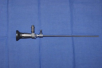
This is a smaller instrument than the flexible cystoscope and is 10mm circumference in size. The visualization is not as good when using this smaller instrument, but when the caliber of the distal urethra is between 10-16 French, the larger scope can’t be used. When the caliber of the urethra is smaller than the scope towards the tip of the penis (distal urethra), we do not attempt urethroscopy because we know that we cannot pass the instrument even a short distance.
Urethroscopy provides a direct look at the appearance along the length of the urethra up to the beginning of the stricture, and also a direct look at the stricture when the urethral stricture is first encountered. However, since the scope cannot be advanced through the stricture without trauma, the exact length of the stricture and the presence or absence of any additional strictures cannot be determined with urethroscopy using s cystoscopy-urethroscope. This highly important additional information is best obtained with X-ray contrast imaging called a retrograde urethrogram (RUG) and voiding cystourethrogram (VCUG).
Overview
The standard procedures for urethral imaging are a retrograde urethrogram (RUG) and a voiding cystourethrogram (VCUG). These are X-ray procedures that we perform in the office, often at the same time as cystoscopy. Films are taken during injection, and during voiding. These studies, when properly performed, will always define the exact location and length of the urethral stricture. It is essential to know this information prior to discussing urethral stricture treatment options, as the treatment decision should be based on first knowing all of the details related to the stricture, not just that there is one!.
Retrograde Urethrogram (RUG)
The retrograde urethrogram (RUG), which is in countries outside the USA also called an ascending urethrogram, is performed with the patient in the oblique position. This means that the patient is tilted 45 degrees. An initial film (called a scout film) is obtained before the actual procedure is performed. This is to confirm that the position and exposure is correct before any contrast is instilled. Then, gauze is gently wrapped around the head of the penis and gentle traction is applied to place the penis on stretch. This is painless. Then, contrast is gently instilled into the urethra using a specialized adaptor developed by Dr. Gelman.
This adaptor, called the Gelman Adaptor, is now commercially available. Dr. Gelman has no financial relationship with the medical device company for this or other inventions, and chose to avoid all industry relationships where there would be monetary compensation to avoid a conflict of interest. This adaptor is now pictured in the major textbook of Urology called Campbell-Walsh Urology.
The adaptor painlessly forms a seal at the tip of the penis. No needles or catheters are required and no balloons are ever inflated in the urethra.
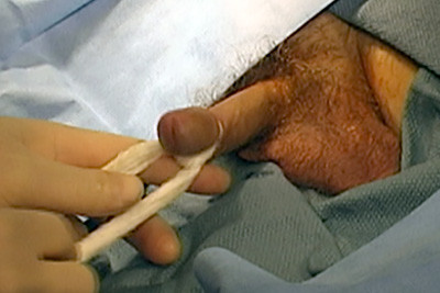
Gauze in place

Contrast slowly instilled
As contrast (clear fluid that looks like water but appears bright white on an X-ray) is injected, a film is obtained. This study permits visualization of the entire urethra. This is an example of a RUG when the anterior urethra is normal.
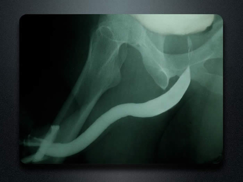
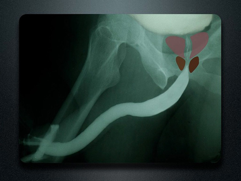
The retrograde urethrogram, also called a RUG, best evaluates the anterior urethra. As previously mentioned, this is the portion of the urethra between the tip of the penis and the beginning of the posterior urethra (the membranous and prostatic urethra and bladder neck). Think about the anterior urethra as a tube or a hose that has nothing to do with leakage. The tube just transports urine. The RUG only provides very limited information about the posterior urethra, the part between the anterior urethra and the bladder. This is because the posterior urethra is not like a hose and has sphincters that open (during urination) and close (all other times). During the injection of contrast, the patient is not voiding, and when a patient is not voiding, the sphincters along the posterior urethra in the area near the prostate are closed, and the prostatic urethra is narrow. It is normal for the posterior urethra to be narrow and pinched off as a RUG is performed. If the external sphincter muscle and prostate were visible on the film, the image would appear as shown below.
Since the posterior urethra may be narrow because of a stricture, or may be narrow because it is normal, you can’t diagnose a stricture of the posterior urethra with a RUG. Therefore, while a RUG is the best imaging test to evaluate the anterior urethra, it is a poor study to evaluate the posterior urethra.
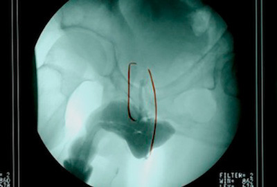
RUG with superimposed graphics demonstrating the prostate and external sphincter (red)
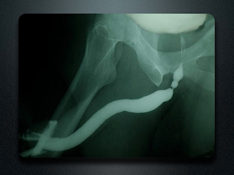
In a patient with a bulbar urethral stricture (bulbar is part of the anterior urethra), contrast passes through the stricture and outlines the area of narrowing and the normal area proximal to the stricture (proximal means on the bladder side of the stricture). Look at how narrow the urethra is at this point. However, since the patient was able to pass urine through the stricture, contrast could be instilled backwards to outline the urethra on both sides of the stricture. As long as there is some opening, since fluid can pass through small spaces, we are able to obtain excellent visualization of the entire urethra, something that can’t be done when only flexible cystoscopy is performed.
Film demonstrating bulbar stricture
Voiding Cystourethrogram (VCUG)
The VCUG is the best study to evaluate the posterior urethra. This is how it is done: As the RUG is performed, some contrast enters the bladder, and this can be seen on the film. The patient is then asked to urinate, and during urination, a film is obtained. It is often the case that patients are unable to void (urinate) after contrast is injected during the RUG as only 60cc of contrast (a small amount) is instilled for this study, and the patient simply may not feel the urge to urinate. Dr. Gelman will then instill additional contrast very, very slowly as to not cause pain until there is an urge for the patient to urinate. During urination, the bladder neck and external sphincters of the posterior urethra are supposed to relax and open as the bladder is contracting. It is a normal reflex. If the posterior urethra is widely patent during voiding, this confirms that this portion of the urethra is normal.
The following is a RUG and VCUG in a patient with a bulbar stricture. Graphics are superimposed (on the VCUG) to show the location of the prostate and external sphincter muscle (posterior urethra). Notice how the posterior urethra is closed off at rest but wide open during urination. The RUG nicely demonstrates the exact length and location and severity of the bulbar stricture, and the VCUG confirms the posterior uretha is normal.
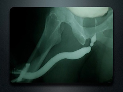
RUG
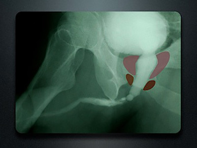
VCUG
However, if there is a narrowing within the posterior urethra during voiding, then this indicates a stricture. In this example, there is a bulbar stricture seen during injection. The posterior urethra is not wide open as would be expected at rest. However during voiding, the area of the membranous urethra remains very narrow.
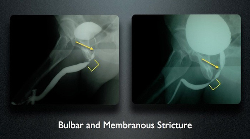
In the above example, if the RUG were the only test performed, then the additional posterior stricture would have been missed. We do not want to risk missing strictures when doing imaging and being “surprised” to find additional strictures at the time of surgery. This could compromise the surgery, and it is for this reason that we perform complete imaging when indicated. Together, the RUG and VCUG provide an evaluation of the entire anterior and posterior urethra. The patient with the 2 separate strictures had a history of dilations and internal stricture incisions, called direct vision internal urethrotomy. Most likely, the stricture was initially only involving the bulbar urethra, and the failed treatments through the penis caused damage to the membranous urethra.
A non-invasive test to measure the flow rate is called a Uroflow. The patient urinates into a funnel shaped collection device in the restroom and this device measures the flow rate over time. This test documents just how fast the urine flows, and is totally non-invasive, but does not tell you the exact internal size of the urethra. The flow rate can vary, depending on how much is in the bladder and other factors. . Another test that can be performed is a bladder scan to check the Post Void Residual (PVR). After urination, an ultrasound probe is placed over the bladder and the bladder can be visualized, and the volume within the bladder can be calculated. The amount that remains in the bladder after urination is called the Post Void Residual or PVR. This does not provide any information about the urethra, but does provide information about possible bladder damage. The bladder should be empty immediately after urination and a high PVR suggests bladder damage. When the bladder empties well, then that is an indicator of good bladder function. Ultrasound and MRI of the urethra are other imaging tests that can be utilized. However, we have generally not found those tests useful.
When patients report having prior urethral imaging, they are encouraged to bring both the films and the reports to their initial visits. If the films are adequate, they do not need to be repeated. However, over the years, we have observed that the vast majority (over 95%) of urethral imaging studies performed at outside facilities are inadequate and do not include both the RUG and VCUG. Often, the reports contain inaccurate interpretations with a misdiagnosis. We are in the process of reviewing our observations and plan to submit our findings for publication given that this appears to be such a pervasive problem. Most General Urologists and Urologists who do not exclusively specialize in male urethral and penile surgery often do not personally perform urethral imaging. It is not practical for them to have X-ray equipment in their offices given that only a very small percentage of their practices are patients with strictures. We have observed that most patients are referred to Imaging Centers for their diagnostic urethral imaging. These Radiology Imaging Centers perform many diagnostic imaging tests such as chest X-rays, CT scans, abdominal ultrasounds etc. and rarely perform urethral imaging.
We have observed that the majority of outside studies are performed using a technique where, instead of the use of the Gelman Adaptor, a catheter is advanced into the urethra a short distance, and a balloon is inflated to create a seal. This is a technique we never use. One problem is that some strictures are towards the tip of the penis and a catheter cannot be advanced even a short distance without creating trauma to the stricture. In addition, when the balloon is inflated in the urethra to create a seal , this painfully dilates and can tear and damage normal urethra. We have used calipers to measure the French size (circumference) of a catheter balloon, and observed that inflation of a catheter balloon with only 2cc of air or water is associated with a balloon caliber of 50 French. A normal urethra is only approximately 24 French in the fossa and around 30 French in the penile portion. These balloons are generally inflated in the fossa navicularis (the part of the urethra closest to the tip of the penis, causing dilation and often very painful tearing of the urethra in this area.
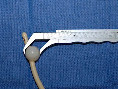
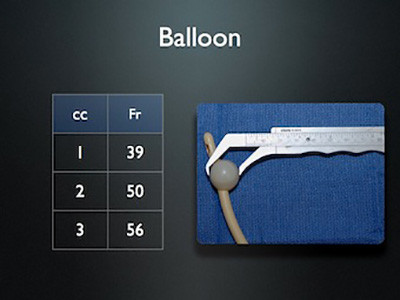
We have actually seen patients referred for strictures in the bulbar urethra (urethral stricture in the part of the urethra under the scrotum far from the tip of the penis) who previously underwent imaging using a balloon technique, and then developed a separate stricture where the catheter was inflated towards the tip of the penis. These patients then require reconstruction of both strictures. After the balloon is inflated, any traction on the catheter to place the penis on stretch can lead to the catheter being pulled out. Therefore, films are often obtained with the penis not on stretch, and the stricture length is underestimated. The following is an example of an outside study using a balloon inflated in the urethra with the penis not on stretch. The impression was that the stricture was 2cm in length. We repeated the study with the penis on stretch using our standard technique, and the stricture was actually 5 cm in length.
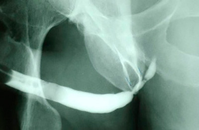
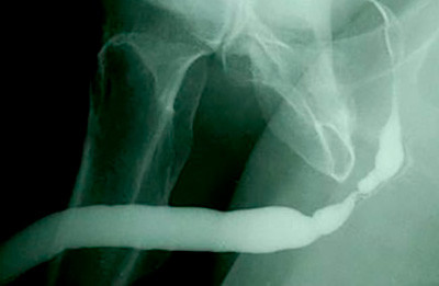
We have seen a number of operative dictations from failed surgeries where it was indicated that during the surgery, it was discovered that the stricture length was longer than anticipated. In these cases, proper imaging would have accurately predicted the stricture length and location, allowing the proper procedure to be planned.
For example, when the position is not sufficiently oblique, the urethra is seen “on end”, preventing proper assessment of the stricture length. If someone is showing you a pencil, and the tip of the pencil is facing you and the rest of the rest of the pencil is facing away from you, it is hard to know the length of the pencil. However, if you see the pencil from the side, you can see both ends and everything in between, and you can tell the length. Same with urethral imaging.
This is the example of an outside film that suggested a 2 cm obliteration of the urethra. The patient was flat on the table with the film taken front to back. We repeated the study with the patient in the proper oblique position (turned partially sideways with the penis on stretch to the side, and the actual length of obliteration was 8 cm.
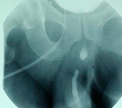
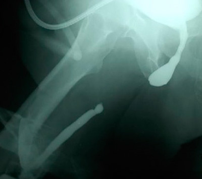
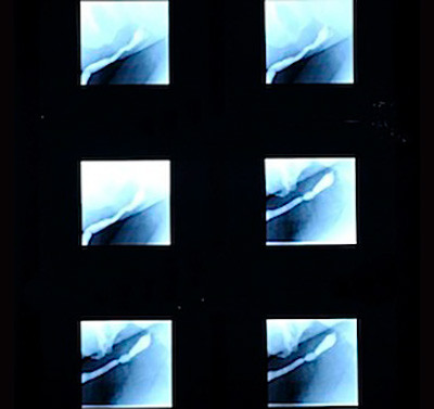
Almost all outside studies we see are performed using fluoroscopy, an imaging technique that allows many films to be taken in a short period of time. This offers the advantage of instant gratification where you can take images in real time, like a movie. The problem with most fluoroscopic studies is that round low quality images are produced with a very small field of view. Often, only a portion of the urethra is seen. We prefer 1 good urethral image over 70 bad images. The following are outside images in comparison to the repeat urethral X-ray imaging performed at our center. Notice how the 1 good image clearly shows the location and length of the area of narrowing.
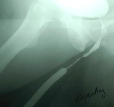
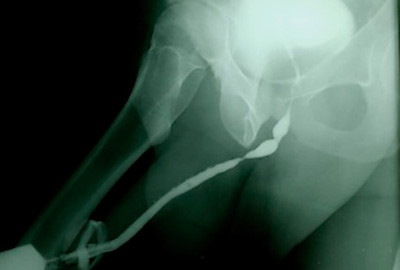
In some cases, the physicians and technicians attempt to fill the bladder by advancing a catheter into the bladder to instill contrast. However, if a catheter could be easily advanced into the bladder, then that would indicate there is no stricture. Since patients do have urethral strictures, this approach to urethral stricture diagnostic testing generally traumatizes the urethra. This is a fluoroscopic image obtained after an attempt was made by the radiologist to advance a catheter through the stricture to then inject contrast. This caused a urethral tear, and the contrast then leaked into the surrounding tissues. This patient was then referred to our center and the study was repeated without the use of a catheter as shown. There was no need to catheterize or cause damage, and we were able to clearly identify the urethral stricture.
Dr. Gelman strongly believes that it is best that Urologists who perform urethral reconstructive surgery personally perform or at least supervise high quality proper urethral imaging procedures, as accurate maging is necessary to provide a specific diagnosis and formulate a correct treatment plan.
At the Center for Reconstructive Urology, the majority of our patients used to be referred by their local urologists for stricture evaluation and treatment. However, with increasing frequency, patients are self-referred as they are not happy with the care they are obtaining locally, and seek care at our Center. A referral is not required. In many cases when a patient is seen by a doctor for the first time, the visit is only a consultation and any tests that are indicated are then scheduled for another time. This is not our practice or preferred approach.
All patients who seek care at the Center for Reconstructive Urology are contacted by Dr. Gelman. We determine if an evaluation at the time of consultation is appropriate. When indicated, we prefer to perform all of the above diagnostic procedures at the time of initial consultation, as the information provided permits an immediate definitive and specific urethral stricture diagnosis. Our patients can then be given definitive treatment recommendations. Patients have travelled to our Center from 46 different states and 27 different countries, and we know that by seeing and examining a patient alone, we can’t provide definitive urethral stricture diagnosis and treatment recommendation.
All testing is performed in a procedure room adjacent to the consultation room at UC, Irvine Medical Center, and Dr. Gelman personally performs all of the imaging studies. Since 1998, over 3,000 urethral imaging studies have been performed at our Center. We have state-of-the-art imaging facilities that produce high resolution images that are saved as digital files, and also printed on high resolution 14 x 17 inch radiographic film and high resolution print images. One copy is placed in the chart, and when patients are referred, a copy is sent to the referring physician (when the patient is referred). Films are printed at the time of the procedure and then reviewed with the patient.
Once we see and examine a patient and then perform a complete urethral stricture diagnostic evaluation, at the same visit, we can then discuss all options and the risks and expected outcome of each option. It is frequently the case that men with a long history of urethral stricture disease are for the first time shown high definition imaging of the urethra and made aware of all options. There is no obligation for patients to then schedule surgery. However, if that is the plan, we can also compete all of the necessary paperwork at the time of the initial visit to facilitate surgery scheduling.

