Call today to consult or
make an appointment

Our Approach & Peyronie’s Disease Surgery Results

Our approach to Peyronie’s Disease surgery is to first determine that the disease is stable (no change in curvature for 6 months) and that the erectile function is good.  We then discuss all options.  Surgery is generally the most effective option to straighten the penis.

The curvature correction options are Peyronie’s disease graft surgery or Peyronie’s disease plication surgery. We offer both options, and carefully counsel patients before surgery.  It is not uncommon for men to contact us requesting a particular Peyronie’s surgery, but many of these men are not aware of all of the risks and benefits of each option.  This understanding should come before decision making.

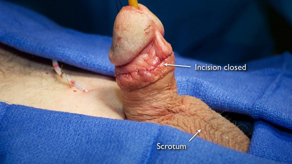
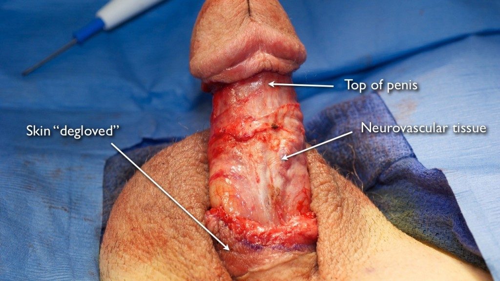
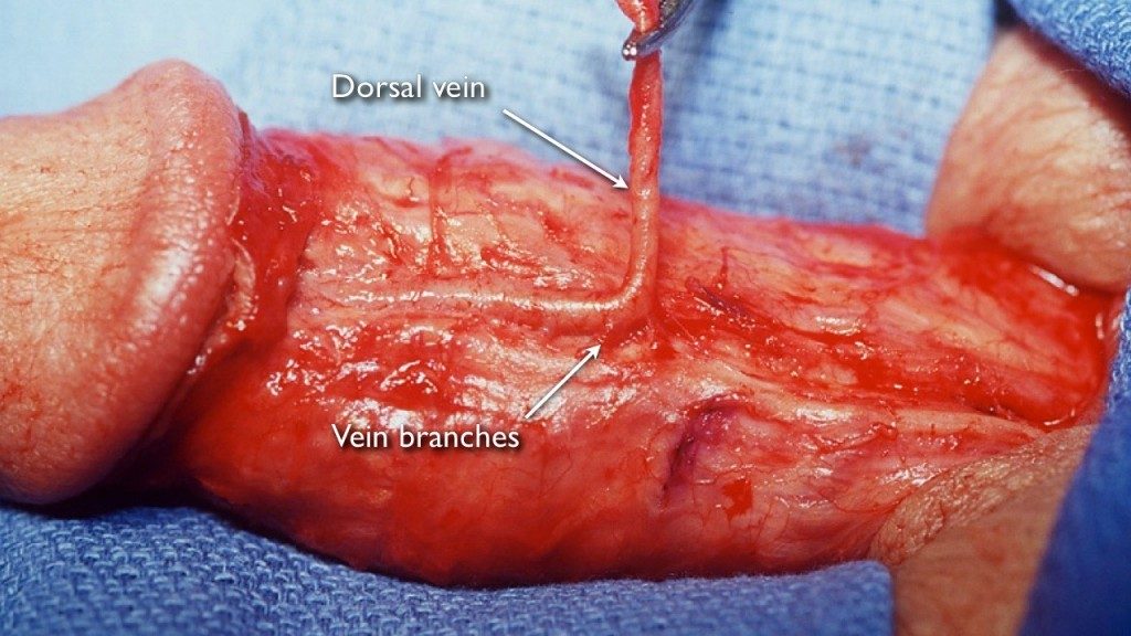
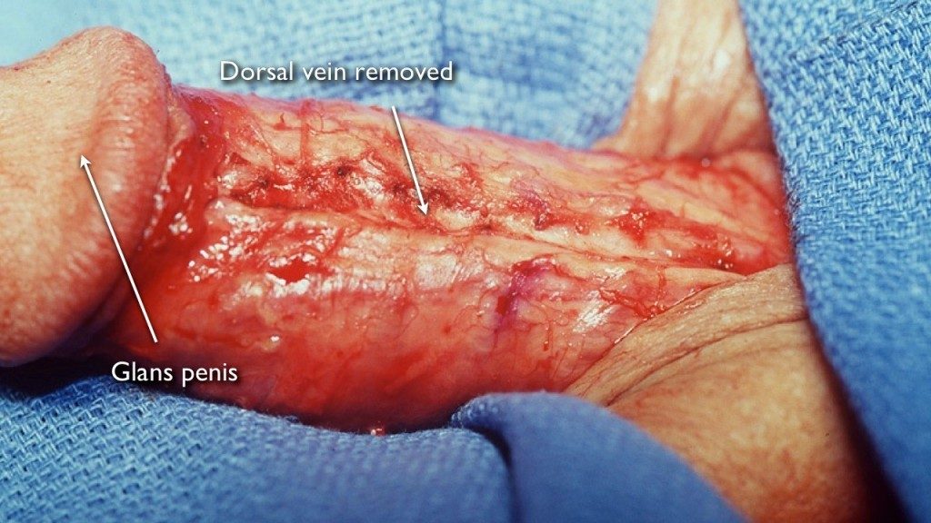
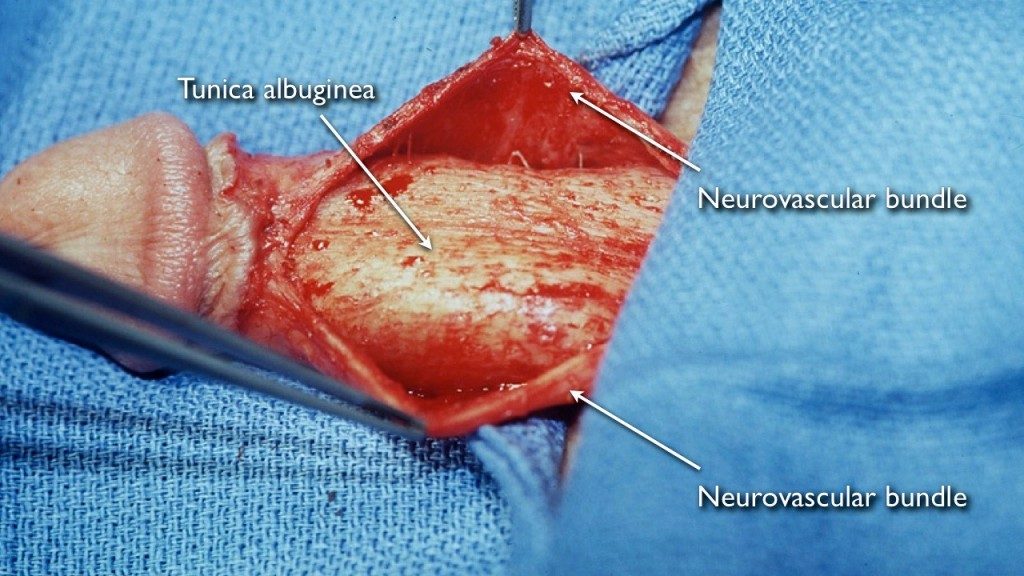
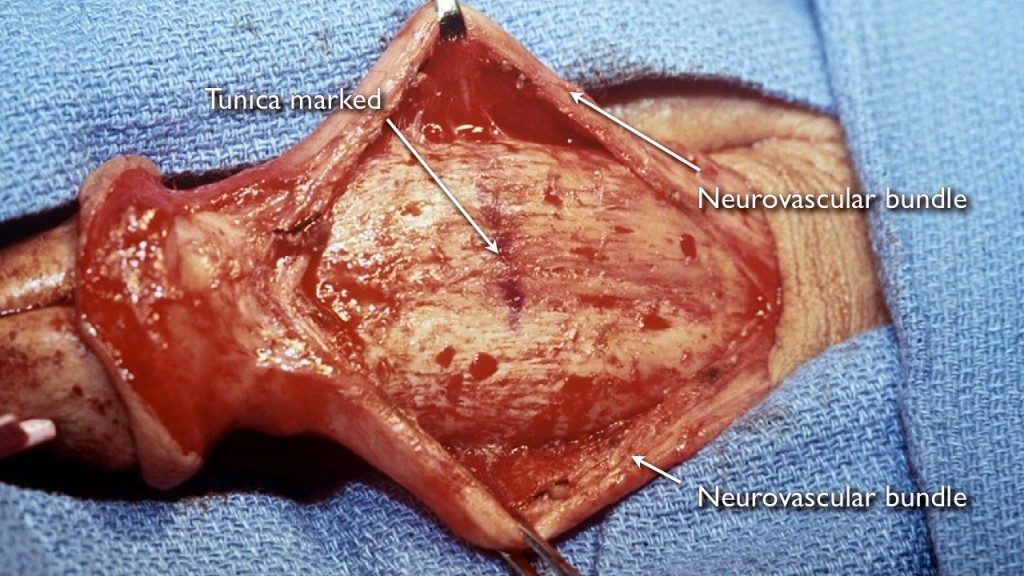
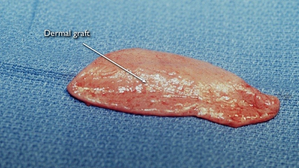
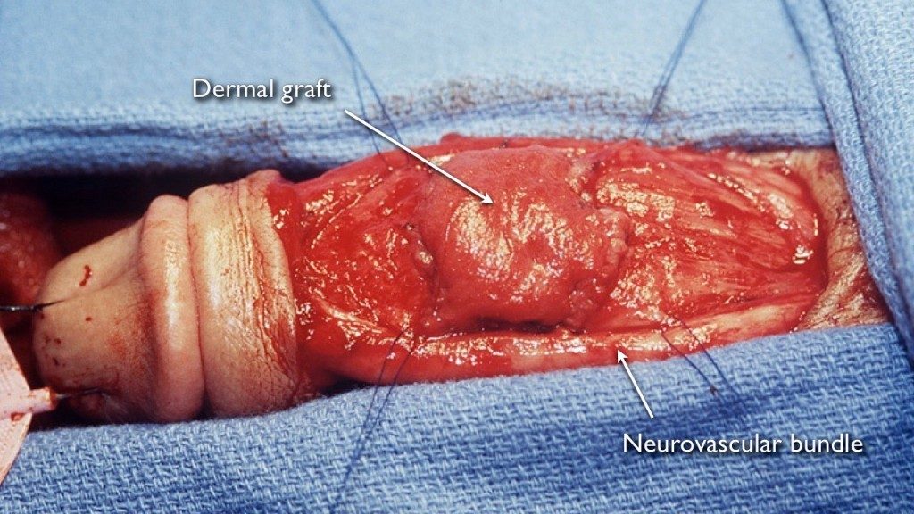
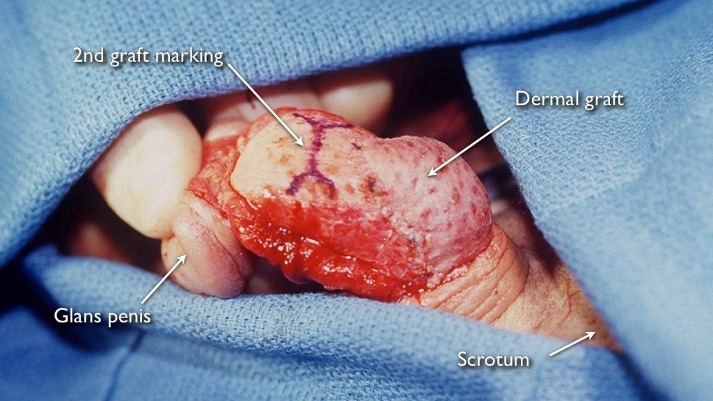
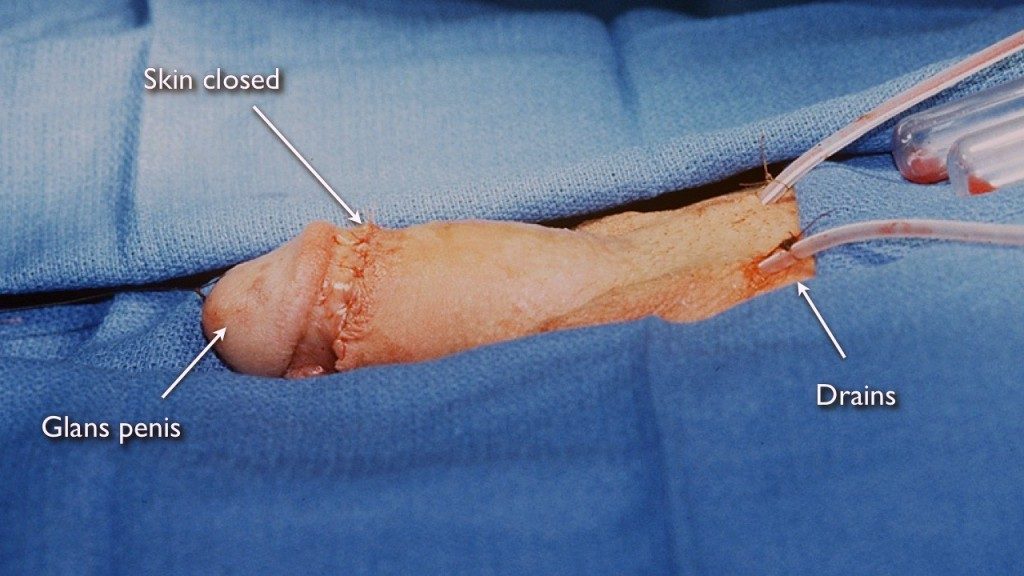
Below we detail our surgical approach and the Peyronie’s disease surgery results for both methods.  These are slideshows depicting actual surgery with graphic pictures demonstrating how we fix penile curvature with before and after photographs.

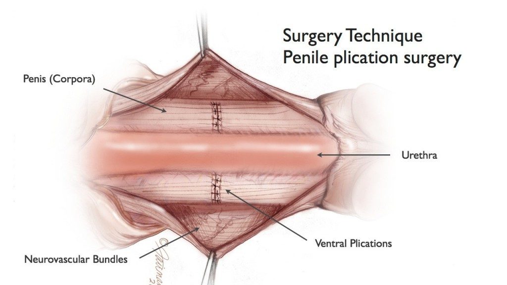
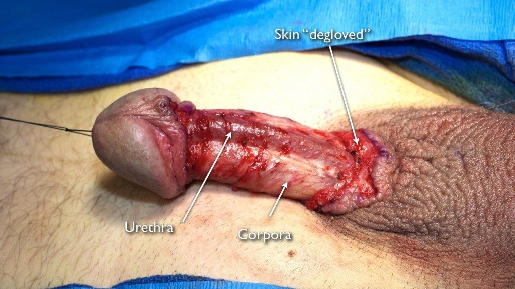
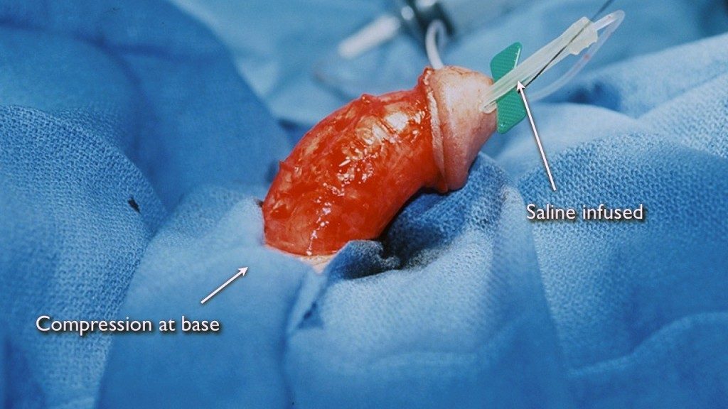
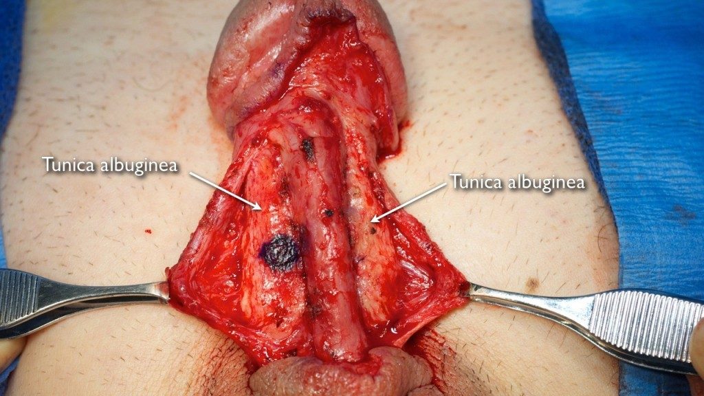
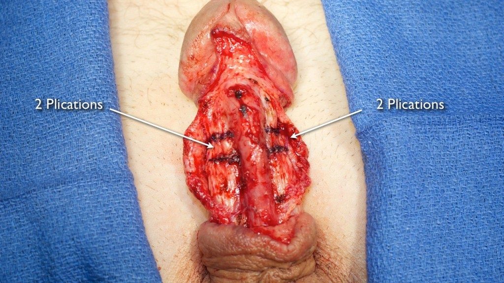
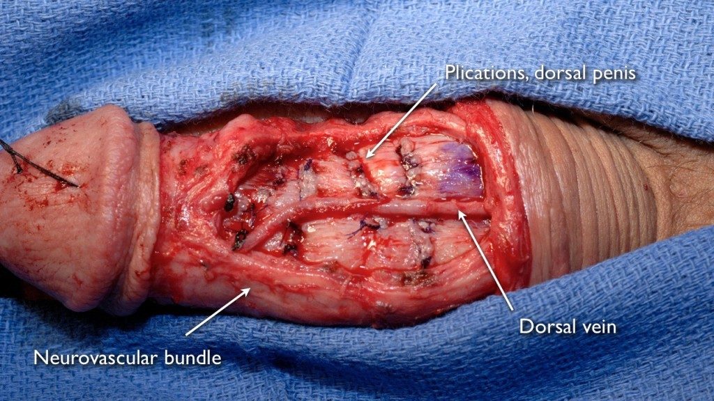
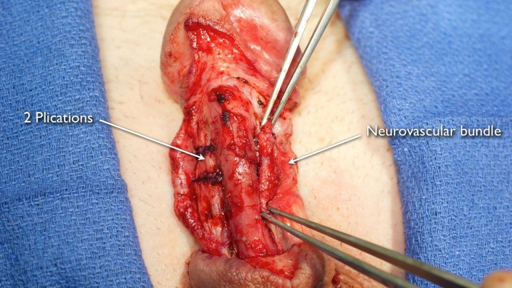
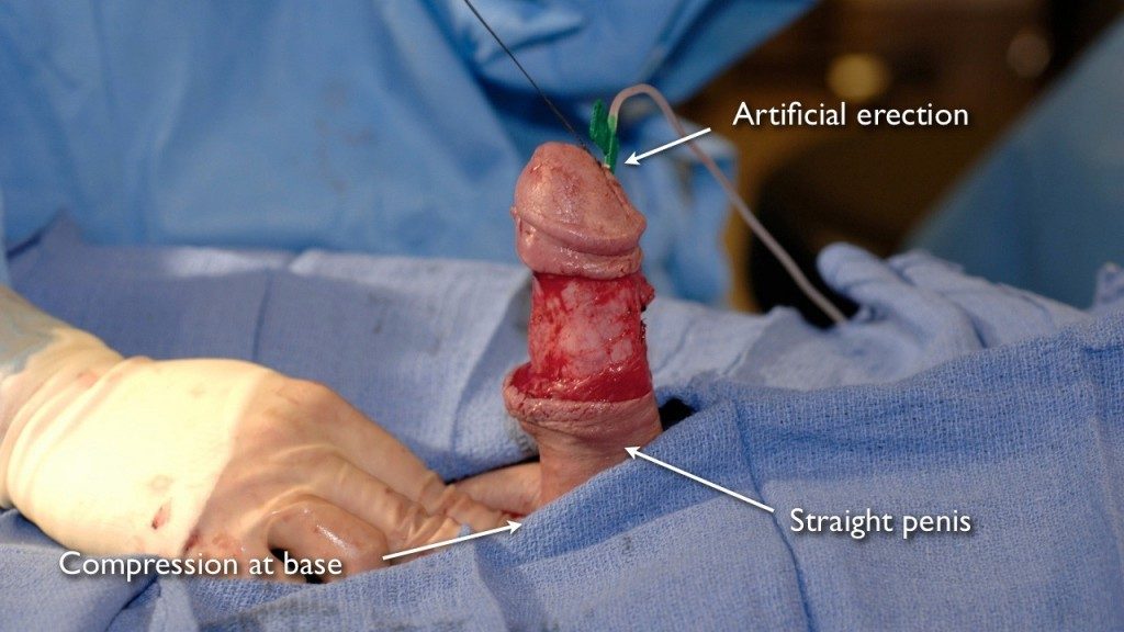
Peyronie’s Disease Plication Surgery

Slideshow of Penile Plication

Slideshow of Penile Plication

The following slideshow demonstrates the technique of correcting penile curvature with Peyronie’s disease plication surgery. In general, this method provides excellent Peyronie’s disease surgery results, with the surgery correcting penile curvature.

Preparing for the Skin Incision

Preparing for the Skin Incision

Peyronie’s disease plication surgery begins with a skin incision. In the picture above, the skin is marked in the location that the incision will take place. The marking is made circumferentially, approximately 1 cm below the area where the head of the penis (glans penis) meets the shaft (coronal sulcus).

The Peyronie’s Disease Fix Incision

The Peyronie’s Disease Fix Incision

The next step is to make the incision, as pictured above. This is called a circumcising incision, even though an actual circumcision (removal of the foreskin) is not performed with this incision. Peyronie’s disease plication surgery is performed under general anesthesia to prevent any discomfort for the patient.

The Penile Skin is “Degloved”

The Penile Skin is “Degloved”

After the skin is incised, the penile skin is “degloved” and brought down to the base of the penis in order to get the best possible exposure to help insure the best possible Peyronie’s disease surgery results. The above picture was taken after the skin is degloved, allowing for the urethra (surrounded by a structure called the corpus spongiosum) and the erectile bodies (called the corpora cavernosa) to be seen.

Saline Infusion to Create an “Artificial Erection”

Saline Infusion to Create an “Artificial Erection”

In the above photograph, saline is used to create an “artificial erection”. Saline is rapidly infused into the penis and the base of the penis is compressed, creating an “artificial erection”. This allows for the abnormal area where the penis is crooked to be easily seen. Plications are then performed on the opposite side of the penile curvature. If there is upward penile curvature, then plications will be performed on the undersurface of the penis.

Curved Penis Tunica Exposed

Curved Penis Tunica Exposed

After the penile skin is degloved and the area of penile curvature is identified, the neurovascular structures (that contain nerves to the glans penis) must be moved to the side, exposing the tissue to be plicated to straighten the bent penis. This tissue is called the tunica albuginea. We perform this dissection under optical magnification without the use of a tournequet, and expertise is required to avoid damaging the sensitive nerves.

Penis Plication Marking

Penis Plication Marking

A pen is used to mark the exposed tunica albuginea to be plicated. The removal of the tissue between the yellow lines will shorten the penis in that area by an amount corresponding to the distance between the lines. When there is penile curvature, the long side is shortened to tilt the bent penis straight.

Penile Plication Compare

Penile Plication Compare

In this Peyronie’s Disease surgery photograph, the neurovascular bundles have been mobilized on the right side of the undersurfase of the penis (ventral-lateral) which is on the left side of the picture, and the area to be plicated is marked. This picture demonstrates the comparison of the appearance before and after the mobilization.

Peyronies Tunica before Penile Plication

Peyronies Tunica before Penile Plication

When upward penile curvature is corrected, we plicate along the undersurface of the penis on both sides of the urethra. In this photo, the neurovascular bundles have been lifted off the tunica albuginea. The urethra is in between the areas to be plicated. Curvature could also be corrected by mobilizing the urethra and performing a midline plication. However, it is our preference to not disturb the urethra to best protect the urethra from possible injury.

Penile Test Plication

Penile Test Plication

Before actually making a permanent change to the penis, we first perform a temporary plication using suture to pinch the area. In this photograph of penile plication surgery for Peyronie’s Disease, the suture has been placed but has not yet been tied.

Penile Plication Tied

Penile Plication Tied

After the temporary plication suture is placed, the suture is tied. An artificial erection is then performed to confirm that the plication of this area will help straighten the bent penis. Once confirmed, we remove the temporary suture and perform a permanent plication to correct the penile curvature by performing an oval shaped excision of tissue and a 2 layer closure with absorbable suture.

Peyronies Plication Completed

Peyronies Plication Completed

Plications have been performed with one plication on each side of the urethra along the undersurface of the penis. Our preference is to use absorbable suture that will eventually dissolve so that the patient does not feel the knots of permanent sutures and there is not a risk that a suture will "pop" leading to a recurrence of penile curvature.

Ventral Penile Plications

Ventral Penile Plications

When there is severe penile curvature, multiple plications are needed to fix a curved penis. In this patient where the penis is abnormally bent upwards, there was only partial correction of the curvature after a single plication on each side of the urethra. We then performed one more plication on each side, and this completely straightened the penis.

Congenital Curvature Plication

Congenital Curvature Plication

Another cause of an abnormal penis is Congenital Penile Curvature. Men who report life long curvature, especially to the side, are born with unequal development of the tunica albuginea of the penis. During an erection, one side is abnormally longer than the opposite side, and this causes a bent penis. In this photograph, plication is performed to fix the penile curvature. In general, congenital penile curvature is best managed with plication and medical management options and stretching devices are of no value.

Dorsal Penis Plication

Dorsal Penis Plication

When there is downward penile curvature, which is less common than an upward bent penis, the plications are performed along the top of the penis to tilt the penis back upward. This is an especially delicate dissection as important nerves and arteries are mostly along the top part of the penis. In this patient, 3 plications were required. Prior to surgery, he had disabling 90 degree downward penile curvature. After surgery, his penis was straight, and he was able to resume sexual intercourse.

Peyronies Surgery

Peyronies Surgery

After the plications are all performed and the penis is straight, the neurovascular bundles are then replaced to again cover the tunica albuginea. In this Peyronie’s Disease surgery picture, on one side, the neurovascular tissue is being repositioned to cover the exposed tunica albuginea as it did before the surgery.

Bent Penis Straightened

Bent Penis Straightened

Before the skin is closed following penile plications, a final artificial erection is performed to confirm the fix of the Peyronie's Disease curvature. In this photograph, the artificial erection revealed that the penis is now completely straight.

Peyronies Surgery Complete

Peyronies Surgery Complete

The skin incision here has been closed with absorbable sutures. These sutures do not have to be removed. Eventually, they dissolve. We leave a small tube under the skin for less than 24 hours to drain any blood. However, bleeding after surgery is rare and to date, we have never had the complication of significant bleeding after penile curvature correction surgery.

Peyronie’s Disease Graft Surgery

Slideshow of Penile Grafting

Slideshow of Penile Grafting

These pictures demonstrate the use of graft surgery as a Peyronie’s disease fix. Penile grafting is an effective penile curvature treatment for selected patients that can provide patients with successful Peyronie’s disease surgery results.

The Penis is Degloved to Treat Dorsal Penis Plaque

The Penis is Degloved to Treat Dorsal Penis Plaque

Similar to plication surgery, Peyronie’s disease graft surgery begins with a circumferential skin incision to “deglove” the penis. In the above picture, the dorsal aspect of the penis (top part) is seen. This is typically the area where penile plaque is felt in patients with upward penile curvature.

The Dorsal Vein is Identified

The Dorsal Vein is Identified

The graft will be placed on the tunica albuginea of the penis, a structure located deep in the neurovascular bundles. These bundles provide sensation and blood flow to the glans penis. In order to expose the tunica albuginea and place the graft, the neurovascular bundles must be carefully moved aside. There is a vein that runs along the middle of the top of the penis called the dorsal vein, and this vein must be identified.

The Dorsal Vein is Removed to See the Tunica Albuginea

The Dorsal Vein is Removed to See the Tunica Albuginea

The dorsal vein can be removed without consequence, and this allows the surgeon to see the tunica albuginea along the midline, an area in which there are no neurovascular structures. In this photograph of Peyronie’s Disease graft surgery, the vein is being removed as branches are cauterized or tied.

Dissect Away the Neurovascular Bundles

Dissect Away the Neurovascular Bundles

After the dorsal vein is removed, there is an opening down to the tunica albuginea. This allows the surgeon, using optical magnification, to carefully dissect the adherent neurovascular bundles away from the tunica albuginea. During the dissection, it is essential to avoid cutting through or damaging these sensitive structures.

Tunica Albuginea

Tunica Albuginea

In this Peyronie’s Disease surgery picture, the neurovascular bundles are being dissected away from the tunica albuginea on the right side (top part of the picture). The tunica albuginea contains the penile plaque. Although some surgeons use a “penile tourniquet” to control bleeding during this dissection, we never need to use this device.

Penis Plaque Exposing

Penis Plaque Exposing

The tunica albuginea is further exposed in this surgery photograph with additional dissection on the left side. Graft surgery to straighten the bent penis lengthens the shortened scarred area by adding tissue, which in this case is along the top part of the penis.

Additional Distal Exposure

Additional Distal Exposure

In some men with Peyronie’s Disease, the affected tunica albuginea extends towards the tip of the penis. In this surgery photograph, the neurovascular structures are mobilized all the way to the glans penis itself in preparation for placement of graft tissue to correct the penile curvature.

Graft Area

Graft Area

Once the neurovascular bundles are fully mobilized in the area of maximum penile curvature, which is confirmed with an artificial erection, the tunica albuginia containing the plaque is marked, and will then be incised. One option is to remove the plaque and then graft. However, there is a greater risk of erectile dysfunction with plaque excision, and the plaque is generally only excised when so calcified and hard, the graft cannot be sutured to the affected area.

Dermal Graft

Dermal Graft

There are several different tissues that can be used as graft material during Peyronie’s Disease surgery. This is a picture of a dermal graft harvested from an area of the abdomen called the “love handle”. Dermis is tissue between the skin and the fat. An advantage of this tissue is that it is readily available in a very adequate amount.

Vein Graft

Vein Graft

Another graft material used by surgeons during graft repair to correct the penile curvature associated with Peyronie’s Disease is a vein graft from the dorsal vein of the penis and/or veins of the leg. We do not prefer to use veins as the quantity of material is less limited with dermal grafts.

Dorsal Dermal Graft

Dorsal Dermal Graft

In this Peyronie’s Disease surgery photograph, after incision, a dermal graft has been placed as a patch to lengthen the shortened portion of the penis. After the graft is sutured in place under optical magnification, an artificial erection is performed to assess the extent of curvature correction.

Second Penis Graft to Correct Curvature

Second Penis Graft to Correct Curvature

Although a single dermal graft can often completely straighten an abnormally bent penis in a man with Peyronie’s Disease, in more severe cases, an additional graft may be required to completely correct the penile curvature. In this surgery picture, a graft has been placed along the mid portion of the penis, and upward curvature towards the tip of the penis remains. A second graft area is marked. In this patient, there was complete penile curvature correction after the placement of a second dermal graft.

Wound Closed, Peyronie's Surgery After Picture

Wound Closed, Peyronie's Surgery After Picture

During Peyronie’s Disease surgery, after the grafts are placed to fix the penile curvature, the neurovascular bundles are re-positioned to cover the exposed tunica albuginea and grafts. The skin incision is then closed with absorbable suture after small temporary drains are placed. These drains are removed prior to discharge from the hospital.

Evaluating Candidates for Peyronie's Disease Surgery

We evaluate the patient’s complete medical history and perform a physical exam and view pictures of the erect penis to determine the best treatment method and if and when surgery may be an appropriate option to fix the Peyronie’s curvature. During this process, we educate our patients so they fully understand the cause of their penile curvature and why they may have pain with erections (if they have painful erections). It is important to discuss the risks and potential benefits of each treatment option, as a patient cannot give proper legal informed consent to treatment without knowing all options.

When a patient has Peyronie’s disease that is in the mature phase and has good erectile function, we ask if the curve interferes with sexual relations. Penile curvature can completely prevent penetration, limit possible sexual positions, or make intercourse uncomfortable for the partner of the patient. These are all valid reasons to pursue a surgical Peyronie’s disease fix. Surgery to straighten a curved penis is generally not considered a cosmetic procedure, so it should be covered by insurance (that does not mean the insurance will pay all costs, but should consider it “medically indicated” and covered based on benefits).

When the curve is mild and not disabling, some patients desire surgery because they find the curvature embarrassing and psychologically troublesome. These patients are advised to weigh the risks vs benefits of surgery. If there is no functional impairment, the patient should carefully consider observation.

Peyronie’s Disease Surgery Results

At the Center for Reconstructive Urology, we have a very high Peyronie’s disease surgery success rate and a very low complication rate. Dr. Gelman is fellowship-trained and expert with over 22 years of experience performing surgery to correct penile curvature, leading to favorable Peyronie’s disease surgery results. Both Peyronie’s disease graft surgery and Peyronie’s disease plication surgery are technically challenging procedures.

blank