After a urethral stricture diagnostic evaluation, a urologist will decide which treatment method is best to widen the narrowed portion of the patient’s urethra. There are various different types of treatments, all of which are appropriate in different circumstances and situations. Depending on the symptoms exhibited and the results of the diagnostic evaluation, a patient may be treated with a urethral stricture dilation, or an internal urethrotomy (DVIU). In the past, Urethral stents (called the Urolume®, made by AMS – American Medical Systems) were a treatment option, but these were found to be a poor option and taken off the market.
Urethral stricture dilation is a common treatment method for smaller strictures that measure one centimeter or less in length. The urethral stricture dilation treatment consists of a urologist progressively stretching the stricture using a series of dilators that gradually increase in size. Depending on the type of stricture, different types of dilator instruments will be utilized. For distal strictures (towards the tip of the penis), larger metal instruments called urethral sounds are used for dilation. More specifically, one of the most commonly used urethral sounds are called Van Buren urethral sounds, pictured below. Another common dilation method involves the use of filiforms and followers, which can also be seen below.
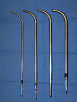
Van Buren Sounds
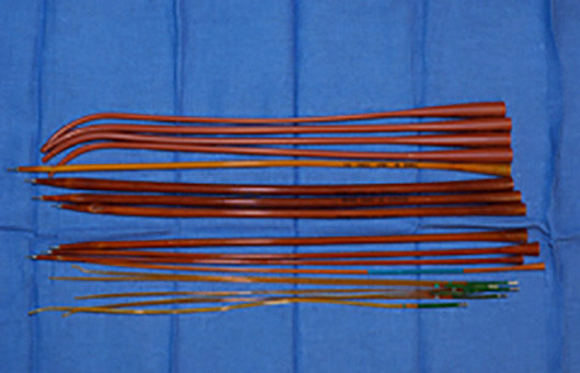
Filliforms and Followers
The filiform is a long and very narrow instrument that is advanced through the urethral stricture to aid in the dilation process. Once the filiform is advanced easily through the stricture, it is attached to progressively larger dilating instruments. These dilating instruments are then carefully guided through the stricture, providing progressive dilation.
Another stricture dilation method is called balloon dilation. This technique involves a specialist gently advancing a flexible tip guidewire through the stricture. Once the guidewire has reached the stricture, a balloon dilating catheter is advanced over the guidewire, and the balloon is guided to the area of narrowing. In the past the balloon had to be guided as a blind procedure, meaning that the specialist had no visibility of the inside of the urethra as they attempted to treat the stricture. However, in 1998 Dr. Gelman developed a balloon dilating catheter, now commercially available (Cook Urological and Boston Scientific – called the Uromax), that allows the stricture to be dilated under direct vision. This incredible innovation has made balloon dilation easier and safer than other dilation methods, leading to the device being published on the cover of the Journal of Endourology in 2011. Performing the procedure under direct vision may be associated with a lower complication rate, as blind dilation methods can result in complications such as false passage and urethral perforation.
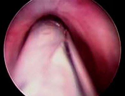
Unopened as Seen Through a Scope
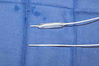
Balloon Dilator Closeup
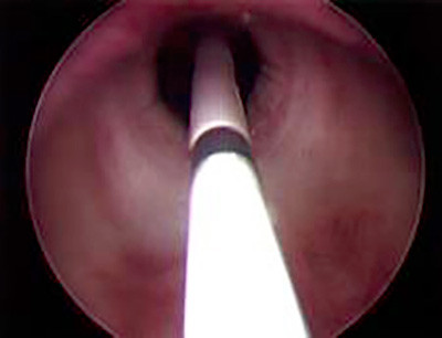
Opened as Seen Through a Scope
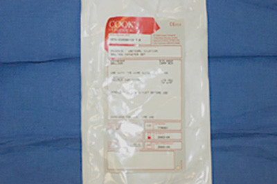
Balloon Dilator Package
Dr. Gelman has no commercial interest in this or other devices and has never accepted royalties to avoid any conflict of interest.
Once the balloon reaches the area of narrowing, it is slowly inflated. Then the balloon is subsequently deflated, and the dilating catheter and guidewire will be removed. Alternatively, some specialists will keep the guidewire in place and insert a temporary stenting catheter over the guidewire after the balloon is deflated.
After initial dilation, the patient may have a temporary and/or intermittent urethral catheter placed to prevent the urethra from immediately narrowing again. In some cases, following urethral dilation, a patient may be asked to perform self-catheterization. Self-catheterization is a procedure where the patient will periodically insert a catheter (tube) into his urethra in an attempt to prevent stricture recurrence, using the catheter as a dilating instrument.
An internal urethrotomy is a surgical procedure that will internally incise the stricture. The procedure is performed in the operating room using an endoscopic instrument, which is a small telescope that is advanced through the penis. Since internal urethrotomy is performed using a telescope, it is often referred to as a DVIU, which stands for direct vision internal urethrotomy. Other names for the procedure include an optical urethrotomy or visual urethrotomy.
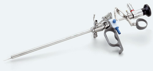
The DVIU procedure begins with the endoscopic instrument inserted into the penis. Near the tip of the instrument is a small blade that can be deployed once the stricture is reached, cutting the stricture internally to “open it up” in one or more places. Once the stricture is opened, an indwelling catheter is placed to keep the urethra open while the urethra heals, which usually takes somewhere between 3 and 5 days.
Some patients are under the false impression that this procedure “cleans out” and removes scar tissue. However, the urethra does not have a build-up of scar tissue inside that can be scraped out. Instead, with urethral stricture disease, the urethra itself is narrow.
After the catheter is placed, the best outcome would be for cells to grow into the open wound so that when the catheter is removed, the urethra remains at a healthy width. This is called epithelialization. Unfortunately, what usually happens is something called wound contraction. Wound contraction is when the body responds to the incision by forming a contracted scar, which can lead to recurrent strictures. The reality is that patients treated with multiple incisions can oftentimes find the procedure complicated by recurrence.
The Urolume® urethral stent involved the placement of an internal metallic stent that had the appearance of a circular chain link fence, as shown below. The objective of the stent was to help open the narrow stricture area of the urethra to widen the narrow area and “stent it open”.
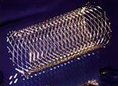
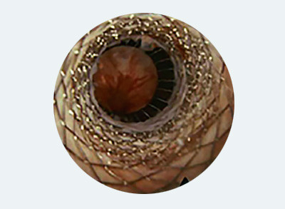
The urethral stent was placed into the urethra endoscopically (through the penis) after the stricture was incised. The objective was for the stent to stop the urethra from contracting as scarring again occurs, preventing the recurrence of strictures. As the lining of the urethra heals, the concept was that it would eventually cover the stent, keeping the stent in place permanently. While this procedure was technically simple to perform, it was unfortunately associated with a high failure and complication rate, as strictures often formed despite the presence of the urethral stent.
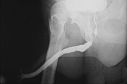
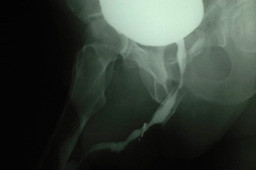
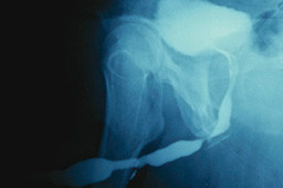
Although the urethral stent was once thought to be an excellent treatment option for recurrent bulbar urethral strictures, it has been shown to compare very unfavorably to open repair. When patients developed strictures after a urethral stent has been placed, the repair was considerably more challenging than it would have been otherwise. These complications resulted in the Urolome urethral stent being taken off of the market, and it is no longer available.
At UC Irvine, we have successfully reconstructed the urethra of many patients referred to our institution after stent failures, and our results were published in the Journal of Urology in 2007. All of our patients have had a technically successful outcome following open repair, making it the best option for patients with recurrent urethral strictures after urethral stent placement.
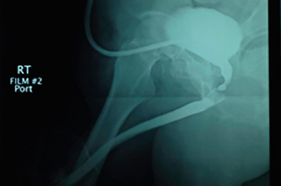
Before
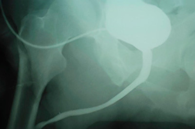
After
In general, patients diagnosed with urethral strictures are often given the impression that they “need” a dilation or urethrotomy (DVIU) to treat their strictures. These are options, but for the patient to make the right decision, he needs to be aware of all options. Unless the length of the stricture is known from urethral imaging (retrograde urethrogram, not just a look with a scope up to where the stricture begins), one can not truly be informed of the best options. Dilation and DVIU are an option that offers the potential for cure, but the cure rate is very low. The cure rate is not 50% plus, and for strictures longer than 1.5-2 cm and for recurrent strictures, the cure rate is close to if not 0. This is because when the urethra is cut, it is injured on purpose. It is lacerated internally. When the urethra is dilated or stretched, it is often torn. When there is bleeding after a dilation, that would indicate an injury rather than the simple stretching of something elastic. When the urethra is cut or dilated-torn open, the urethra is wider at that moment in time. However, it then heals. When it heals, it heals with a scar formation. That is supposed to happen. Unfortunately, most of the time, the urethra then again becomes narrow. When this procedure is repeated, there can be an expected build-up of scar tissue, often making the stricture longer and more dense. Repeated dilations and DVIUs should be considered a management tool that offer the advantage of simplicity but inferior outcomes to a properly performed urethroplasty. One can always try to “keep the stricture open” or “stablize” the urethral stricture with intermittent catheterization, but this is also a management tool, and patients should not think that if they catheterize themselves long enough, they can then stop some day and the urethra will stay open indefinitely.

